
 3 months ago
A project called Antscan has generated high resolution images of thousands of ants, representing over 700 species. To make it happen, researchers brought preserved ants from collections around the world to a particle accelerator in Germany. There, a powerful synchrotron x-ray source combined with a vial-swapping robot allowed the researchers to build a collection of 3D ant images, inside and out. Each voxel (like a 3D pixel) has a resolution of 1.22 micrometers—enough to see the tiny hairs on ant bodies, and distinguish individual muscle fibers.
Antscan researcher Julian Katzke joins us to describe the background of the project, and how the images could be used for science and art.
Check out images from the Antscan projet:
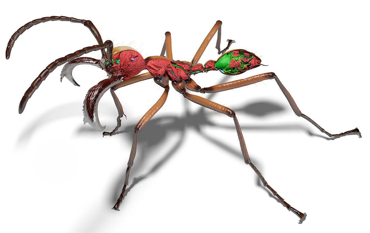 A 3D render of the army ant Eciton hamatum, based on x-ray images from the Antscan project, provides a high-resolution view of the insect’s exoskeleton and organs. Credit: Thomas van de Kamp
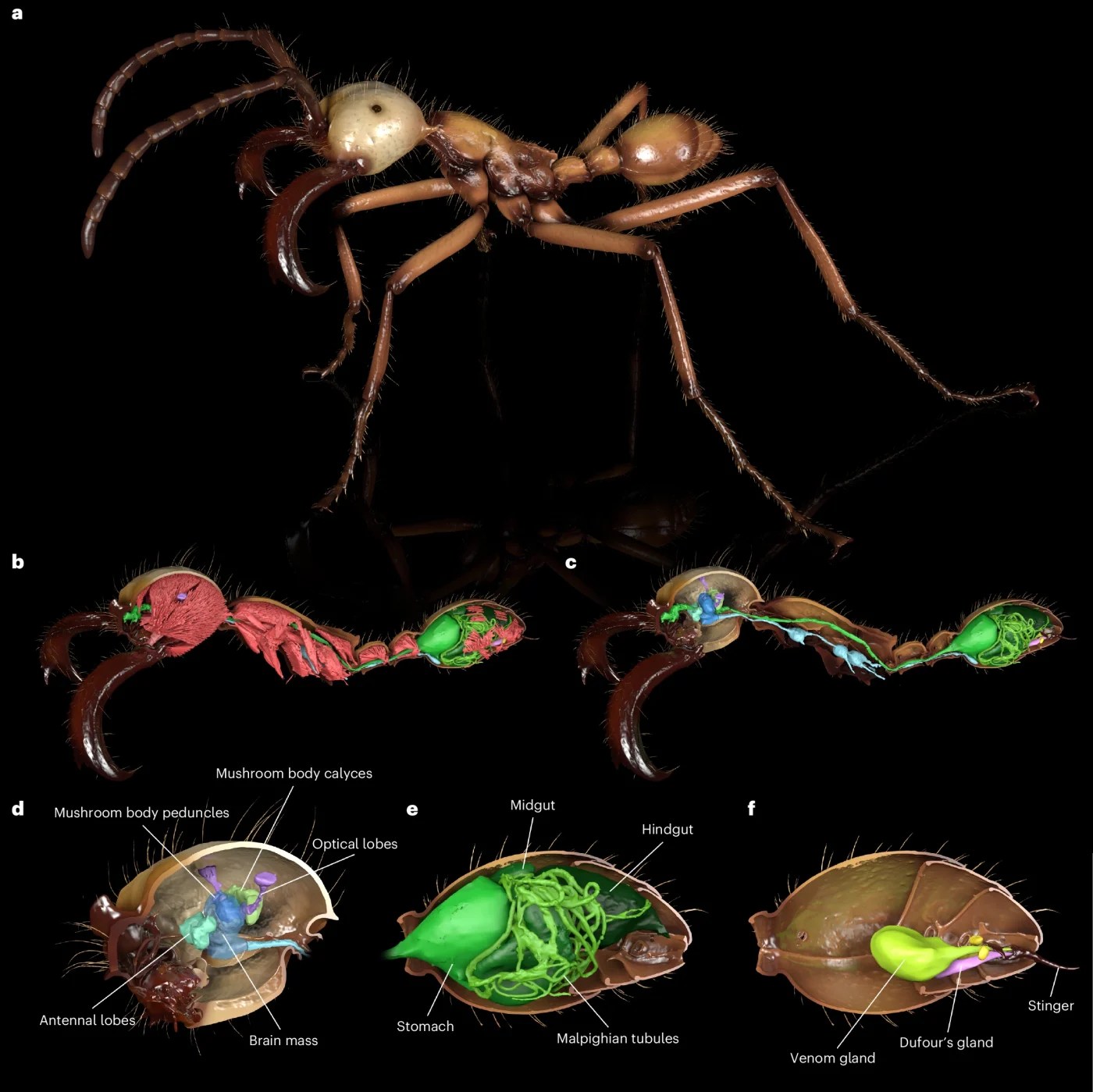 Renderings show the segmented cuticle and tissues representative of the level of detail captured with synchrotron micro-CT. a, Full habitus of the ant with an animated, more life-like pose and colors inspired by photographs. b, Cuticle cut at the sagittal section revealing internal tissues with muscles in red occupying most of the internal space in an ant’s body. c, Removing the muscles reveals the digestive tract (green) and the nervous system (blue). d–f, Zoomed-in renderings focusing on the ant brain (d), gut (e) and sting apparatus (f), respectively. Credit: Nature
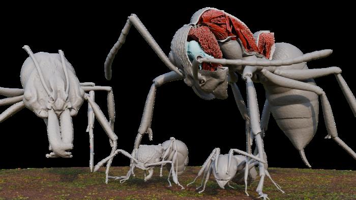 Four Okinawan ants reproduced from Antscan data: Odontomachus kuroiwae (large left), Diacamma cf. indicum (large right), Pristomyrmex punctatus (small left), Technomyrmex brunneus (small right). Diacamma is shown with a portion of its exoskeleton removed, revealing internal organs like part of its nervous system (blue) and muscle fibers (red). Credit: Julian Katzke
Donate To Science Friday
Invest in quality science journalism by making a donation to Science Friday.
Donate
Segment Guests
Julian Katzke
Dr. Julian Katzke is a postdoc at the Smithsonian National Museum of Natural History. He worked on the AntScan project while a PhD student at the Okinawa Institute of Science and Technology.
Segment Transcript
The transcript is being processed. It will be available 2-3 days after this story’s publication date.
Meet the Producers and Host
About Flora Lichtman
Flora Lichtman is a host of Science Friday. In a previous life, she lived on a research ship where apertivi were served on the top deck, hoisted there via pulley by the ship’s chef.
About Charles Bergquist
As Science Friday’s director and senior producer, Charles Bergquist channels the chaos of a live production studio into something sounding like a radio program. Favorite topics include planetary sciences, chemistry, materials, and shiny things with blinking lights.


